AN INTERPROFESSIONAL APPROACH TO KIDNEY STONE PREVENTION AND MANAGEMENT
Faculty:
L. Austin Fredrickson, MD, FACP
L. Austin Fredrickson is an Associate Professor of Internal Medicine at Northeast Ohio Medical University, where he serves as core faculty and teaches diagnostics, therapeutics, clinical skills, and health humanities. He is board-certified in general internal medicine and practices rural primary care.
Liz Fredrickson, PharmD, BCPS
Liz Fredrickson, PharmD, BCPS, is an Associate Professor of Pharmacy Practice and Pharmaceutical Sciences at the Northeast Ohio Medical University (NEOMED) College of Pharmacy, where she is course director of the Parenteral Products and Basic Pharmaceutics Lab courses.
Pamela Sardo, PharmD, BS
Pamela Sardo, PharmD, BS, is a freelance medical writer and licensed pharmacist. She is the founder and principal at Sardo Solutions in Texas. Pam received her BS from the University of Connecticut and her PharmD from the University of Rhode Island. Pam’s career spans many years in retail, clinics, hospitals, long-term care, Veterans Affairs, and managed health care responsibilities across a broad range of therapeutic classes and disease states.
Topic Overview
The development of renal calculi, or kidney stones, is a common and recurrent condition affecting millions worldwide and significantly impacts quality of life, productivity, and healthcare utilization. Patients may present asymptomatically or with acute renal colic, hematuria, and/or nausea and vomiting. If left untreated, nephrolithiasis can lead to urinary obstruction, infection, and long-term kidney damage. Accurate diagnosis through urinalysis and imaging is essential to confirm the presence, size, and location of stones and to guide management decisions. This interprofessional educational activity reviews key guideline recommendations, emphasizing the evolving approach to acute and preventive management. Learners will explore evidence-based strategies for initial symptom control, medical expulsive therapy, and indications for urologic consultation, as well as key components of metabolic evaluation following stone passage. The program also highlights pharmacologic and lifestyle interventions to reduce the risk of recurrence. Participants will gain a comprehensive understanding of stone prevention and management principles, the healthcare team’s role in medication counseling and adherence support, and the value of interdisciplinary collaboration in optimizing long-term renal health and patient outcomes.
Accreditation Statements
In support of improving patient care, RxCe.com LLC is jointly accredited by the Accreditation CouncilTM for Continuing Medical Education (ACCME®), the Accreditation Council for Pharmacy Education (ACPE®), and the American Nurses Credentialing Center (ANCC®), to provide continuing education for the healthcare team.


This activity was planned by and for the healthcare team, and learners will receive 2 Interprofessional Continuing Education (IPCE) credits for learning and change.
Joint Universal Activity Number: The Joint Accreditation Universal Activity Numbers assigned to this activity are as follows:
Pharmacists: JA4008424-0000-26-014-H01-P
Pharmacy Technicians: JA4008424-0000-26-014-H01-T
Credits: 2 contact hour(s) (0.2 CEU(s)) of continuing education credit.
Credit Types:
IPCE Credits - 2 Credits
AAPA Category 1 Credit™️ - 2 Credits
AMA PRA Category 1 Credit™️ - 2 Credits
Pharmacy - 2 Credits
Type of Activity: Knowledge and Application
Media: Computer-Based Training (i.e., online courses)
Estimated time to complete activity: 2 contact hour(s) (0.2 CEU(s)), including Course Test and course evaluation.
Release Date: January 30, 2026 Expiration Date: January 30, 2029
Target Audience: This educational activity is for Physicians, Physician Assistants, Pharmacists, and Pharmacy Technicians
How to Earn Credit: From January 30, 2026, through January 30, 2029, participants must:
Read the “learning objectives” and “author and planning team disclosures;”
Take the “Educational Activity Pre-Test;”
Study the section entitled “Educational Activity;” and
Complete the Educational Activity Post-Test and Evaluation. The Educational Activity Post-Test will be graded automatically. Following successful completion of the Educational Activity Post-Test with a score of 70% or higher, a statement of participation will be made available immediately. (No partial credit will be given.)
CME Credit: Credit for this course will be uploaded to CPE Monitor® for pharmacists. Physicians may receive AMA PRA Category 1 Credit™️ and use these credits toward Maintenance of Certification (MOC) requirements. Physician Assistants may earn AAPA Category 1 CME credit, reportable through PA Portfolio. All learners shall verify their individual licensing board’s specific requirements and eligibility criteria.
Statement of Need
Patients presenting with kidney stones are a common, recurrent occurrence, and increasingly associated with obesity, diabetes, and aging. Despite the frequency, healthcare professionals underutilize guideline-directed strategies for diagnosis, management, and prevention. Avoidable pain, complications, and costs are the result. Disparities in timely diagnosis, analgesia, and follow-up disproportionately affect Black patients and those with lower socioeconomic status, contributing to recurrence and lower quality of life. It is important to keep up to date with evolving evidence, such as changes in the role of thiazide diuretics. As expanded use of citrate and uric acid-lowering treatments is discussed within interprofessional teams, opportunities exist to review gaps between current practice and the American Urological Association (AUA) guidelines and the European Association of Urology (EAU) recommendations. This activity aims to strengthen recognition of risk factors, apply evidence-based imaging, prescribe treatments that expel stones, and optimize pain control. Shared decision-making and a team approach to care can increase lifestyle changes and optimize pharmacologic prevention plans.
Learning Objectives: Upon completion of this educational activity, participants should be able to:
Recall guideline recommendations for kidney stone evaluation and prevention
Compare and contrast pharmacologic and lifestyle interventions to prevent recurrence
Explain interprofessional collaboration in patient education and long-term care
Evaluate patient-specific risk factors for the development of kidney stones
Disclosures
The following individuals were involved in developing this activity: Austin Fredrickson, MD, FACP, Liz Fredrickson, PharmD, BCPS, and Pamela M. Sardo, PharmD, BS. None of the individuals involved in developing this activity has a conflict of interest or financial relationships regarding the subject matter. There are no financial relationships or commercial or financial support relevant to this activity to report or disclose by RxCe.com or any of the individuals involved in the development of this activity.
© RxCe.com LLC 2026: All rights reserved. No reproduction of all or part of any content herein is allowed without the prior, written permission of RxCe.com LLC.
Educational Activity Pre-Test
Which of the following recommendations aligns with current kidney stone prevention guidelines?
Restrict all dietary calcium to lower urinary calcium
Increase fluid intake to achieve ≥2.5 L urine/day
Avoid all dietary oxalate completely
Use thiazides routinely for any calcium stone former
Which lifestyle modification most effectively reduces the risk of stone recurrence?
Avoiding all dairy products
Increasing fluid intake to achieve urine SG <1.010
Following a high-protein diet
Avoiding all citrate-containing foods
Which pharmacologic therapy is most appropriate for uric-acid stone prevention with persistent low urine pH?
Allopurinol alone
Thiazide diuretics
Indapamide
Potassium citrate
Educational Activity
An Interprofessional Approach to Kidney Stone Prevention and Management
Introduction
The development of renal calculi, or kidney stones, is a common and recurrent condition affecting millions worldwide and significantly impacts quality of life, productivity, and healthcare utilization. Patients may present asymptomatically or with acute renal colic, hematuria, and/or nausea and vomiting. If left untreated, nephrolithiasis can lead to urinary obstruction, infection, and long-term kidney damage. Accurate diagnosis through urinalysis and imaging is essential to confirm the presence, size, and location of stones and to guide management decisions.
This interprofessional educational activity reviews key guideline recommendations, emphasizing the evolving approach to both acute and preventive management. Learners will explore evidence-based strategies for initial symptom control, medical expulsive therapy, and indications for urologic consultation, as well as key components of metabolic evaluation following stone passage. The program also highlights pharmacologic and lifestyle interventions to reduce the risk of recurrence. Participants will gain a comprehensive understanding of stone prevention and management principles, the healthcare team’s role in medication counseling and adherence support, and the value of interdisciplinary collaboration in optimizing long-term renal health and patient outcomes.
Pause and Ponder
Think about the last patient you encountered with a kidney stone (or imagine one if no one comes to mind):
What challenges did they face, not only with pain, but with diagnosis, follow-up, prevention, and navigating the healthcare system?
Terminology
Urolithiasis and nephrolithiasis are terms often used interchangeably but technically refer to distinct aspects of urinary stone disease.1 Nephrolithiasis is specific to the formation and presence of stones within the kidneys, and encompasses the intricate process of stone development within the renal parenchyma and renal collecting system.1,2 This is detailed in Figure 1 below.2 Urolithiasis is a broader definition that includes not only nephrolithiasis but also the formation or occurrence of urinary stones throughout the entire urinary tract, including the ureters and bladder.1 It is important to differentiate between these terms, as they inform clinical management and treatment approaches.
Nephrolithiasis often requires specific interventions for stones lodged within the kidneys, and urolithiasis requires a more comprehensive evaluation and management of stones throughout the urinary system.1 Most stones still arise from the kidney or ‘start’ as nephrolithiasis, but migrate into other areas. Colloquially, “kidney stones”, known medically as “renal calculi”, comprise true nephrolithiasis but generally any urolithiasis, even if lodged in the bladder or a “bladder stone”.1 For example, a bladder stone may be removed with cystoscopic retrieval, whereas a stone wedged in the kidney itself may require lithotripsy or stent insertion more proximally to the bladder.
Figure 1
Kidney Anatomy2

Epidemiology and Etiology
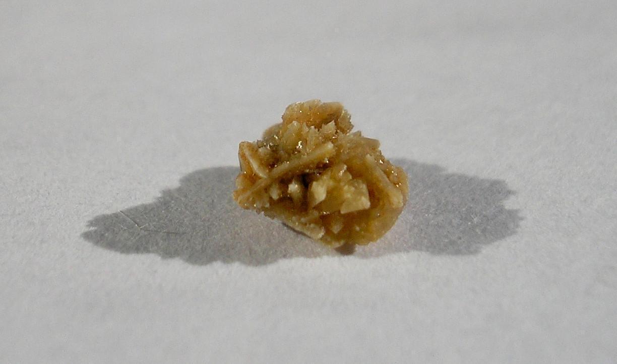
There are various types of kidney stones, and identification will inform a patient’s prognosis, treatment, and preventive care regimen.3 Stones can be a mix of different crystal types, for example, a combination of calcium oxalate and calcium phosphate.3 Stones can also be made up of medications, such as acyclovir, though this is rare. Types of kidney stones are listed in Table 1.4 Figure 2 visually depicts a kidney stone.4
The prevalence of urolithiasis varies globally, with rates estimated between 1-13%.5 The incidence of urolithiasis within the United States (US) has increased in recent years, with an estimated lifetime risk of 10-15%.6
Numerous factors contributed to this rise, including dietary habits, increased body mass index, comorbidities, climate change, and socio-economic conditions.3,5 Recent data suggest approximately 19% of men and 9% of women will develop a kidney stone at least once in their lifetime.3 For men, the peak incidence occurs around 40 years of age and then starts to decrease.3 For women, the peak incidence occurs at 30 years of age.3
Table 1
Types of Kidney Stones3
| Stone Type | Percentage |
|---|---|
| Calcium oxalate | 75% |
| Calcium phosphate | 15% |
| Uric acid | 8% |
| Struvite | 1% |
| Cystine | <1% |
Figure 2
Kidney Stone4
The disease prevalence among Hispanics (6.4%) and blacks (4.3%) has been estimated to be lower than among non-Hispanic whites. However, minority patients have been found to have poorer quality of life, delays in diagnosis and treatment, and a greater incidence of disease recurrence.7 As an example, studies have shown that Black patients receive poorer analgesic care compared to white patients, and one study found Black patients received less analgesia when presenting with nephrolithiasis compared to white patients.8 It is vital that care teams recognize and address these disparities.
In addition to causing disease-related complications and issues for patients, nephrolithiasis is also associated with a significant economic burden for patients due to loss of work and costs related to treatments and surgeries.3 These issues and discrepancies described above also occur for patients with lower socio-economic status.3
Risk Factors
The risk factors for nephrolithiasis can be grouped into four broad categories: dietary, nondietary, urinary, and genetic.3 Diet plays an important role in both the generation and recurrence of kidney stones, with many factors involved in the pathophysiology of nephrolithiasis affected by diet.3 Dietary factors associated with an increased risk of nephrolithiasis include animal protein, oxalate, sodium, sucrose, and fructose intake.3 Intake of calcium, potassium, and phytate is associated with a decreased risk of developing nephrolithiasis.3 Increases in dietary calcium may reduce risk due to a reduction in the intestinal absorption of dietary oxalate, which leads to a decrease in urine oxalate and, thus, a decreased risk of calcium oxalate stones.3 A summary of dietary factors is presented in Table 2.
Table 2
Dietary factors and risk of kidney stone development3
| Dietary Factor | Modification | Potential Stone Risk |
|---|---|---|
| Fluid intake | Reduction | Increased urine saturation/ concentration |
| Sodium intake | Increase | Increased urine calcium and reduced citrate expression |
| Calcium intake | Reduction | Increased urinary oxalate excretion |
| Meat intake | Increase | Low urine pH, increased urine calcium, and reduced citrate excretion |
| Diet content in oxalate foods | Increase | Increased urinary oxalate excretion |
Non-dietary risk factors include age, race, body size, medication use, and the environment.3 While nephrolithiasis can occur in patients ranging from young infants to elderly individuals, it is most common among middle-aged white men.3 Additionally, kidney stone development is most common within the southeastern region of the US.3
The development of kidney stones is also associated with weight gain, and the increase in nephrolithiasis has mirrored the rise in obesity rates in the US.3 Environmental risk factors include those that reduce urine volume, such as limited water or bathroom access.3 Numerous medications are also associated with an increased risk of kidney stone formation. These are listed in Table 3.3
Table 3
Medications Associated with Kidney Stone Formation
| Type of medication | Examples |
|---|---|
| Antibiotics | Ampicillin, amoxicillin, ceftriaxone, sulfonamides |
| Carbonic anhydrase inhibitors | Acetazolamide, topiramate |
| Diuretics | Furosemide, triamterene |
| Laxatives | Overuse of any laxative that results in electrolyte loss |
| Potassium channel blockers | Amiodarone, dalfampridine, sotalol, |
| Reverse transcriptase inhibitors and protease inhibitors | Efavirenz, indinavir, nelfinavir, raltegravir |
| Sulfonylureas | Therapies for type 2 diabetes |
| Others | Aluminum magnesium hydroxide, ascorbic acid, calcium, dexamethasone, guaifenesin, phenytoin, vitamin D |
| GLP-1 Agonists | Case reports |
These medications either promote crystallization in the urine, alter urinary chemistry, decrease mineral solubility, dehydrate the body, or promote urinary stasis.3
Antibiotics can increase the risk of crystal deposition, including through increased urine concentration.
Carbonic anhydrase inhibitors can cause metabolic acidosis by increasing bicarbonate excretion. This can increase the excretion of calcium and uric acid in the urine.
Diuretics cause dehydration and increase concentration, as do laxatives.
The effect of potassium channel blockers on electrolyte homeostasis can predispose to stones.
Certain antiviral medications can also cause metabolic changes that increase the risk.
Sulfonylureas increase the risk of obesity and insulin resistance, both of which can increase the risk of calculi.
Other medications that affect urine composition (namely, calcium or uric acid) or other metabolic disturbances can affect stone formation.
Urinary risk factors also contribute to the development of kidney stones.3 One major urinary risk factor is low urine volume.3 This is considered a modifiable risk factor, with studies highlighting the utility of increasing fluid intake to reduce the risk of nephrolithiasis.3 The pH of urine can also affect the solubility of different types of crystals.3 For example, uric acid stones can only form at a pH <5.5, and calcium phosphate stones at a pH >/6.5.3 Calcium oxalate stone development is not affected by urinary pH. The remaining urinary risk factors are summarized in Table 4.
Table 4
Urinary Risk Factors3
| Risk Factor | Description |
|---|---|
| Urine calcium | Higher urinary calcium excretion increases the risk of calcium oxalate and calcium phosphate stones |
| Urine oxalate | Higher urine oxalate excretion increases the risk of calcium oxalate stone formation |
| Urine citrate | Lower urine citrate excretion increases the risk of kidney stone formation |
| Urine uric acid | High levels of urine uric acid increase the risk of uric acid stone formation |
Genetic factors also play important roles.3 An individual with a family history of kidney stone development is twice as likely to develop nephrolithiasis as someone without a family history.3 To date, two rare monogenic disorders lead to kidney stone development: primary hyperoxaluria and cystinuria.3 Primary hyperoxaluria is an autosomal recessive disorder that leads to excessive endogenous oxalate being created by the liver.3 This eventually causes calcium oxalate stone formation and deposition.3 Cystinuria is also an autosomal recessive disorder.3 This disorder causes the abnormal reabsorption of filtered basic amino acids. Because cystine is poorly soluble, the excessive urinary cysteine excretion leads to cysteine stone formation.3
Active Learning
Match each item in Column A with the correct risk-factor category in Column B.
| Column A – Risk Factor Examples | Column B – Categories |
|---|---|
| 1. High sodium intake | A. Dietary Risk Factor |
| 2. Low urine volume | B. Non-Dietary Risk Factor |
| 3. Primary hyperoxaluria | C. Urinary (Metabolic) Risk Factor |
| 4. High animal-protein diet | D. Genetic Risk Factor |
| 5. Obesity/weight gain | |
| 6. Low urine citrate |
Pathophysiology
The pathophysiology of stone formation is important for effective treatment and management. Understanding the pathophysiology of stone formation also helps the clinician identify risk factors for nephrolithiasis.
The pathophysiology of nephrolithiasis begins with the unique composition of urine, a complex solution often supersaturated with minerals that can crystallize. Under normal conditions, stone formation is prevented by crystallization inhibitors, most notably urine citrate, which binds to calcium to reduce supersaturation, inhibits calcium crystal growth, and enhances the activity of macromolecules that prevent calcium oxalate aggregation.3 When these protective mechanisms are disrupted, mineral deposition begins deep within the kidney.3 Our current understanding finds that stone formation typically originates at the thin limb of the loop of Henle, where calcium phosphate precipitates and accumulates within the renal interstitium.3
These deposits, known as Randall’s plaques, gradually enlarge and eventually erode through the papillary epithelium to become exposed to urine.3 Once exposed, the plaques serve as anchoring sites on which calcium oxalate and calcium phosphate crystals adhere, aggregate, and ultimately form clinically detectable stones.3 This stepwise process often occurs years before symptoms arise and underscores the importance of early identification and modification of metabolic risk factors.3
Case
A 47-year-old man presents for follow-up after passing a 6 mm distal ureteral stone. The stone analysis shows calcium oxalate monohydrate. His 24-hour urine collection reveals:
Urine volume: 1.4 L/day
Urine calcium: 260 mg/day
Urine citrate: Low
Urine oxalate: Normal
His diet includes frequent restaurant meals, processed foods, and minimal fruits/vegetables.
He asks, “What should I change so this doesn’t happen again?”
Patient Presentation and Diagnosis
Patients with nephrolithiasis typically present with acute flank pain, often severe and colicky, and radiating toward the groin as the stone migrates.3 Associated symptoms include nausea, vomiting, urinary urgency or frequency, dysuria, and occasionally gross or microscopic hematuria.3 While classic renal colic is common, many patients, especially older adults, women, and those with comorbidities, may have atypical or vague symptoms, underscoring the importance of maintaining clinical suspicion.11 If patients have experienced a previous episode of kidney stones, these symptoms can assist in making the diagnosis.11 Patients generally present with either renal colic or painless gross hematuria when an acute stone event occurs.3 The pain associated with renal colic can vary in intensity and can increase rapidly. 3
Evaluation begins with a focused physical exam and point-of-care urinalysis, which help detect hematuria and identify signs of a urinary tract infection.3 It can also assist clinicians with differential diagnosis (Table 5). The absence of hematuria does not exclude nephrolithiasis, particularly after several days of symptoms.3 The American Urological Association (AUA) continues to recommend low-dose noncontrast CT of the abdomen/pelvis as the most accurate initial test for diagnosing kidney stones in nonpregnant adults due to its superior sensitivity and ability to identify alternative diagnoses.12 However, in order to minimize cumulative radiation exposure, the European Association of Urology (EAU) recommends an “ultrasound-first” approach in stable patients, reserving low-dose CT for cases in which ultrasound is inconclusive.13 Ultrasound remains the preferred modality during pregnancy and in children. If an ultrasound suggests hydronephrosis without visualization of a stone, a follow-up low-dose CT may still be needed to guide management.
Table 5
Differential Diagnosis of Kidney Stones11
| Clinical Clues | Suggested Diagnosis |
|---|---|
| Dysuria | Interstitial cystitis, prostatitis, urinary tract infection, vaginitis |
| Fever, chills | Nonspecific response to infection or inflammation |
| Hematuria | Benign prostatic hyperplasia, renal glomerular disease, urinary tract infection, urinary tumor |
| Nausea, vomiting | Gastrointestinal disease, intestinal or urinary obstruction, nonspecific response to pain |
| Abdominal pain and tenderness | Acute mesenteric ischemia, cholecystitis, gastrointestinal disease |
| Flank pain and tenderness | Dysmenorrhea, herpes zoster, and pyelonephritis |
| Urinary frequency | Benign prostatic hyperplasia, bladder spasms, high fluid intake, hyperglycemia, urinary tract infection |
Because stones frequently do travel down the urinary tract, the entire tract can be visualized with a plain radiograph of the abdomen.11 This X-ray test is commonly called a KUB, which stands for kidneys, ureters, and bladder.11 Radiopaque stones (namely those with calcium) can be visualized through this modality.11 However, radiolucent stones (such as uric acid stones) may not be visualizable. A more detailed assessment of the soft tissues can be obtained with the CT urogram; however, it also entails increased radiation and cost.11
Figure 3
Algorithm for the Diagnosis and Management of
Acute Kidney Stones11
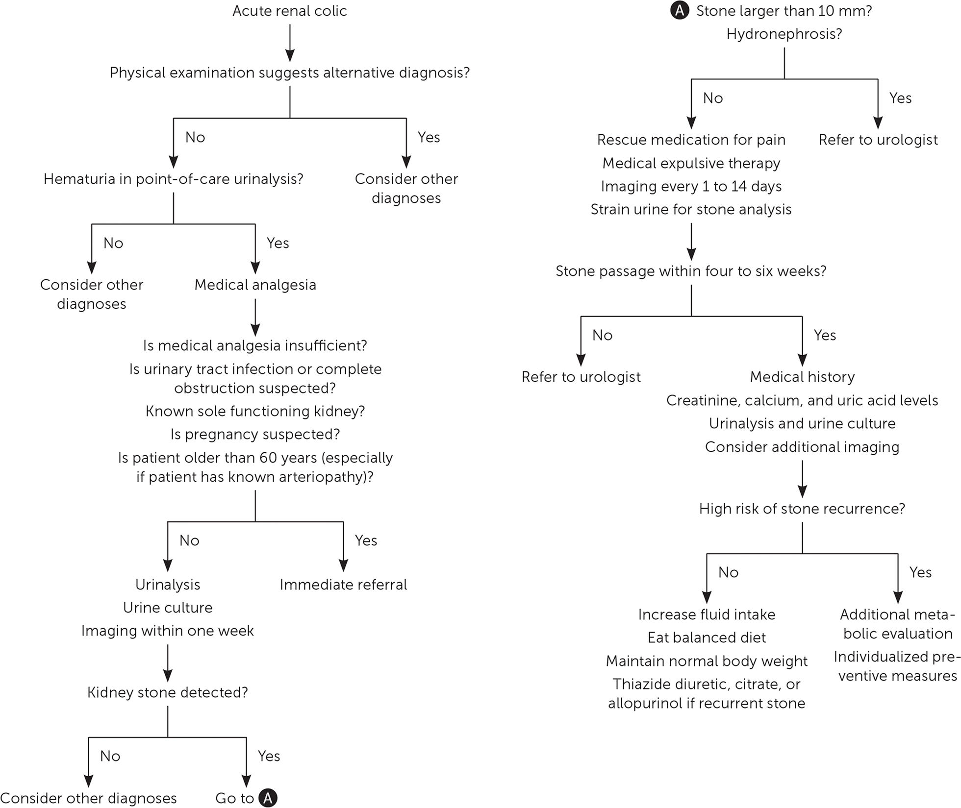
A diagnostic pathway is detailed in Figure 3 above.11 If a stone is found to be larger than 10mm in size or is causing obstructive features of hydronephrosis or kidney injury, it is generally recommended to refer the patient to a urologist. Smaller stones may still be unable to pass; however, even stones as small as 4mm may not pass without intervention.11
Approaches to Treatment
The acute management of kidney stones involves appropriately managing the patient’s pain.11 If not contraindicated, it is recommended to utilize nonsteroidal anti-inflammatory drugs (NSAIDs) over opioids, assuming there is no kidney injury that could be exacerbated, as NSAIDs have greater efficacy and a lower risk of adverse effects.11 Suggested agents include ketorolac at 30 mg or 60 mg intramuscularly.11 If opioids are utilized, it is recommended to avoid meperidine due to the risk of nausea and vomiting associated with this agent.11 If a patient’s pain is uncontrollable, referral to a urologist or emergency department is recommended.11 Patients should also seek emergency care when sepsis is suspected, if they are anuric, if they have a urinary tract infection with renal obstruction, for women who are pregnant or could possibly be pregnant, and in patients with comorbidities or those greater than 60 years of age.11
If referral to a urologist is unnecessary, patients can be managed conservatively.11 For these outpatients, proper hydration is encouraged. Pain medications should be prescribed as needed, and follow-up imaging should be scheduled within two weeks to determine the stone's position and monitor for hydronephrosis.11 If the stone does not pass on its own, the patient should be referred to a urologist for removal.11 It is estimated that approximately 86% of stones pass spontaneously.12
Medical expulsive therapy (MET) uses alpha-blockers to facilitate stone passage by relaxing ureteral smooth muscle, reducing spasms, and improving ureteral dilation. Current AUA and 2024–2025 EAU recommendations support MET only for distal ureteral stones measuring 5–10 mm, the group most likely to benefit.12,13 Alpha-blockers are not effective for stones <5 mm (which typically pass spontaneously), stones >10 mm, proximal ureteral stones, or in pregnant patients. Tamsulosin 0.4 mg daily remains the most widely studied agent, while silodosin may provide slightly higher expulsion rates but carries a greater risk of retrograde ejaculation. Nifedipine is no longer recommended for MET.12,13 Increasing fluid intake or adding corticosteroids does not enhance stone passage and should not be used for this purpose. Stone position and symptoms should be reassessed within 4 weeks. If there is a lack of progression, worsening hydronephrosis, or uncontrolled pain, this warrants referral to urology.
Clinicians should instruct patients to strain their urine to catch the stone. The stone should be sent in a clean, dry container for stone analysis.11 It is recommended to analyze recurrent stones if they have different compositions.11 Once the stone composition is known, appropriately targeted preventative measures can be taken to guard against recurrence.11 Creatine, ionized calcium, and uric acid levels should be measured. Parathyroid hormone levels can also be measured if serum calcium levels are high to assess for hyperparathyroidism.11
Special Considerations
Special consideration should be made for asymptomatic kidney stones, children, and pregnant women.
Asymptomatic Kidney Stones
Kidney stones may sometimes be asymptomatic and only found incidentally on imaging.11 Stones can take weeks to months to grow to a clinically detectable size.3 It has been estimated that 10-25% of stones are symptomatic or require some type of intervention.11 For patients without contraindications, conservative management with active surveillance is appropriate. The patient can be referred for stone removal if issues arise, such as obstruction or recurrent infection.3,11
Children
Due to increasing rates of obesity and diabetes in children, nephrolithiasis is becoming more prevalent in the pediatric population.11 Children who develop kidney stones are more likely to have a metabolic, neurologic, or congenital urinary system structural abnormality.11
Pregnant Women
An estimated 75% of stones in pregnant women are made of calcium phosphate.11 Due to the risk to the fetus, both diagnostic and treatment options are limited in this population.11 In some cases, the development of kidney stones may increase the risk of maternal and fetal complications.11
Pause and Ponder In what ways can pharmacy technicians meaningfully reduce preventable emergency department visits for renal colic through workflow, adherence support, and communication practices? |
|---|
Prevention
Non-pharmacologic and pharmacologic strategies are available to prevent recurrent kidney stones. The method for preventing recurrent stones depends on the risk of recurrence. Lifestyle modifications are recommended for those with a low risk of recurrence.11 These include increasing fluid intake to 2.5-3 liters per day, which helps achieve a urine-specific gravity of less than 1.010.11 Beverages consumed should be neutral in pH, and carbonated drinks should be avoided.11 In addition to increased fluids, patients should eat a balanced diet high in fiber and vegetables and with a normal consumption of calcium (1-1.2 grams per day).11 Patients should be counseled to limit sodium consumption to 4-5 grams daily and animal protein to 0.8-1 grams per kilogram daily.11 Obese and overweight patients should aim to achieve a normal body weight by increasing physical activity.11
Certain medications and supplements can also help prevent recurrent kidney stones. These include thiazide diuretics, allopurinol, and citrate. These agents are typically used when lifestyle modifications are insufficient to prevent stone recurrence.11 The role of thiazide diuretics in preventing recurrent nephrolithiasis has shifted significantly following the NOSTONE Trial (NEJM 2023), which found no statistically significant reduction in stone recurrence with hydrochlorothiazide compared with placebo.14 As a result, thiazides are no longer recommended as a routine preventive therapy.14 They may still be considered in select patients with documented hypercalciuria, but their overall preventive benefit is likely smaller than previously believed.14 Additionally, higher-dose thiazides, particularly chlorthalidone 25–50 mg, carry increased risks, including hypokalemia, hyponatremia, glucose intolerance, and gout.14 Therefore, thiazide use should be individualized based on metabolic evaluation and patient-specific risk factors rather than broadly applied.14
Allopurinol is a second pharmacologic option, specifically for patients who have hyperuricemia and/or uric acid stones.11 It is dosed at 100 mg once daily, and the dose can be increased to 100 mg three times daily if needed.11 Allopurinol can reduce uric acid production and help dissolve uric acid crystals that have formed.11 Currently, insufficient evidence suggests that combining allopurinol with either a thiazide diuretic or citrate is more effective than allopurinol monotherapy.11
Supplementation with citrate can be useful for calcium stones, uric acid, and cystine stones.11 This agent is recommended at a dose of 5 to 12 grams daily.11 The total dose should be divided into three equal doses taken 30 minutes before a meal or snack.11 Potassium citrate alkalinizes the urine, preventing stones that form in acidic environments, and also inhibits crystal formation by increasing the solubility of stone-forming substances, including calcium and uric acid.11 Its use is associated with gastrointestinal symptoms.11 As with allopurinol, there is no evidence that combination therapy with potassium citrate is more beneficial than monotherapy, though the use of potassium citrate with a thiazide diuretic can correct low potassium levels caused by the diuretic.11
As a less expensive option, patients can use unsweetened lemonade to supplement their citrate intake. This can also help patients increase their fluid intake.11 Citrate can also be taken in by consuming foods that are rich in alkali, such as fruits and vegetables.3 Patients who supplement citrate should have both potassium levels and liver enzymes monitored. Potassium levels should be monitored within 2 weeks of starting a supplement, and then at least every 12 months.3
Emerging Therapies
Specialized therapies are increasingly important for managing rare or refractory forms of nephrolithiasis. Lumasiran, an RNA-interference therapy, is FDA-approved for primary hyperoxaluria type 1 and has been shown to markedly reduce endogenous oxalate production, thereby lowering the risk of recurrent calcium oxalate stone formation and systemic oxalosis.15
Collaborative Care
Effective management of nephrolithiasis increasingly relies on a collaborative, multidisciplinary care model that integrates the expertise of physicians, pharmacists, pharmacy technicians, nurses, dietitians, and urology specialists. Primary care clinicians and emergency providers often serve as first-line diagnosticians, identifying symptoms, ordering imaging, and ruling out complications. Pharmacists play a central role in optimizing medication therapy, counseling on pain management, advising on the appropriate use of medical expulsive therapy, reviewing drug–drug interactions, and supporting long-term prevention strategies such as citrate supplementation or uric acid–lowering therapy. Pharmacy technicians enhance this workflow by assisting with medication reconciliation, ensuring accurate dispensing, supporting adherence strategies, and helping coordinate follow-up care.
Dietitians contribute essential expertise in tailoring dietary interventions, such as sodium restriction, adequate calcium intake, and oxalate reduction, based on stone composition and metabolic risk factors. Urologists provide procedural and surgical expertise for patients with obstructing, infected, or non-passing stones and collaborate on metabolic evaluations for recurrent stone formers. Together, this coordinated approach improves diagnostic accuracy, reduces treatment delays, enhances patient education, and supports long-term prevention, ultimately reducing recurrence rates and improving patient outcomes.
Return to Patient Case
The priority intervention is to increase fluid intake to achieve a urine output of ≥2.5 L/day. Additional evidence-based recommendations include:
Increase citrate intake (potassium citrate or dietary alkali such as fruits/vegetables, or unsweetened lemonade).
Reduce sodium intake to help lower urinary calcium excretion.
Maintain normal dietary calcium intake (1,000–1,200 mg/day) to limit intestinal oxalate absorption.
Thiazides are not routinely recommended following NOSTONE but may be considered only if persistent hypercalciuria remains after lifestyle modification.
Summary
Kidney stones, medically known as renal calculi, are a prevalent and persistent condition that brings about considerable pain and distress, leading to missed work, reduced productivity, and heightened healthcare resource utilization. Despite established diagnostic and treatment approaches for kidney stones (urolithiasis), including proven preventive measures, this challenge persists. Recent research highlights disparities in evaluation and care based on socioeconomic factors and race. Familiarity with medications and methods to prevent and treat these painful and organ-threatening conditions is imperative. Given the rising prevalence and varied causes of kidney stones, effective management and prevention require healthcare professionals to have a comprehensive, up-to-date understanding of this ubiquitous condition.
Answer Key to Active Learning on Page 13 above
Match each item in Column A with the correct risk-factor category in Column B.
| Column A – Risk Factor Examples | Column B – Categories |
|---|---|
| 1. High sodium intake | A. Dietary Risk Factor |
| 2. Low urine volume | B. Non-Dietary Risk Factor |
| 3. Primary hyperoxaluria | C. Urinary (Metabolic) Risk Factor |
| 4. High animal-protein diet | D. Genetic Risk Factor |
| 5. Obesity/weight gain | |
| 6. Low urine citrate |
Answer Key
1 → A (High sodium increases urinary calcium and lowers citrate)
2 → C (Low urine volume increases supersaturation)
3 → D (Primary hyperoxaluria is an autosomal recessive genetic disorder)
4 → A (Animal protein lowers pH, increases calcium, lowers citrate)
5 → B (Obesity is a major non-dietary risk factor)
6 → C (Low urine citrate reduces inhibition of CaOx stone formation)
References
Bishop K, Momah T, Ricks J. Nephrolithiasis. Prim Care. 2020;47(4):661-671. doi:10.1016/j.pop.2020.08.005
Wiki Commons. Kidney Anatomy. https://en.wikipedia.org/wiki/Kidney#/media/File:Blausen_0592_KidneyAnatomy_01.png. Accessed November 13, 2023.
Curhan GC. Nephrolithiasis. In: Loscalzo J, Fauci A, Kasper D, Hauser S, Longo D, Jameson J. eds. Harrison's Principles of Internal Medicine, 21e. McGraw Hill; 2022.
Wiki Commons. Kidney Stone. https://commons.wikimedia.org/wiki/File:Blausen_0592_KidneyAnatomy_01.png. Accessed November 13, 2023.
Lang J, Narendrula A, El-Zawahry A, Sindhwani P, Ekwenna O. Global Trends in Incidence and Burden of Urolithiasis from 1990 to 2019: An Analysis of Global Burden of Disease Study Data. Eur Urol Open Sci. 2022;35:37-46. Published 2022 Jan 3. doi:10.1016/j.euros.2021.10.008
Wilcox CR, Whitehurst LA, Cook P, Somani BK. Kidney stone disease: an update on its management in primary care. Br J Gen Pract. 2020;70(693):205-206. Published 2020 Mar 26. doi:10.3399/bjgp20X709277
Scotland KB, Armas-Phan M, Dominique G, Bayne D. Social Determinants of Kidney Stone Disease: The Impact of Race, Income and Access on Urolithiasis Treatment and Outcomes. Urology. 2022;163:190-195. doi:10.1016/j.urology.2021.08.037
Scales CD, Smith AC, Hanley JM, Saigal CS. Prevalence of kidney stones in the United States. Eur Urol. 2012;62:160–165. 10.1016/j.eururo.2012.03.052.
Caudarella R, Vescini F. Urinary citrate and renal stone disease: the preventive role of alkali citrate treatment. Arch Ital Urol Androl. 2009;81(3):182-187.
Ferraro PM, Bargagli M, Trinchieri A, Gambaro G. Risk of Kidney Stones: Influence of Dietary Factors, Dietary Patterns, and Vegetarian-Vegan Diets. Nutrients. 2020;12(3):779. Published 2020 Mar 15. doi:10.3390/nu12030779
Fontenelle L and Sarti T. Am Fam Physician. 2019;99(8):490-496.
Pearle MS, Goldfarb DS, Assimos DG, et al. Medical Management of Kidney Stones: AUA Guideline. J Urol. 2014;192(2):316-324.
Türk C, Neisius A, Petřík A, et al. EAU Guidelines on Urolithiasis. Arnhem, The Netherlands: European Association of Urology; 2024.
von Unruh GE, Hess B, Knoll T, et al; NOSTONE Trial Investigators.
Hydrochlorothiazide and Prevention of Kidney-Stone Recurrence.
New England Journal of Medicine. 2023;388(9):781-791. doi:10.1056/NEJMoa2211705
Michael M, Groothoff JW, Shasha-Lavsky H, et al. Lumasiran for Advanced Primary Hyperoxaluria Type 1: Phase 3 ILLUMINATE-C Trial. Am J Kidney Dis. 2023;81(2):145-155.e1. doi:10.1053/j.ajkd.2022.05.012
DISCLAIMER
The information provided in this course is general in nature, and it is designed solely to provide participants with continuing education credit(s). This course and materials are not meant to substitute for the independent, professional judgment of any participant regarding that participant’s professional practice, including but not limited to patient assessment, diagnosis, treatment, and/or health management. Medical and pharmacy practices, rules, and laws vary from state to state, and this course does not cover the laws of each state; therefore, participants must consult the laws of their state as they relate to their professional practice.
Healthcare professionals must consult their employer, healthcare facility, hospital, or other organization for guidelines, protocols, and procedures to follow. The information provided in this course does not replace those guidelines, protocols, and procedures, but is for academic purposes only, and this course’s limited purpose is for the completion of continuing education credits.
Participants are advised and acknowledge that information related to medications, their administration, dosing, contraindications, adverse reactions, interactions, warnings, precautions, or accepted uses is constantly changing. Any person taking this course understands that such a person must make an independent review of medication information before any patient assessment, diagnosis, treatment and/or health management. Any discussion of off-label use of any medication, device, or procedure is informational only, and such uses are not endorsed hereby.
Nothing contained in this course represents the opinions, views, judgments, or conclusions of RxCe.com LLC. RxCe.com LLC is not liable or responsible to any person for any inaccuracy, error, or omission with respect to this course or course material.
© RxCe.com LLC 2026: All rights reserved. No reproduction of all or part of any content herein is allowed without the prior, written permission of RxCe.com LLC.
RxCe.com
© RxCe.com LLC 2025: All rights reserved.