
BREAST CANCER TREATMENT: A MULTIDISCIPLINARY APPROACH
Faculty:
L. Austin Fredrickson, MD, FACP
L. Austin Fredrickson is an Associate Professor of Internal Medicine at Northeast Ohio Medical University, where he serves as core faculty and teaches diagnostics, therapeutics, clinical skills, and health humanities. He is board-certified in general internal medicine and practices rural primary care.
Liz Fredrickson, PharmD, BCPS
Liz Fredrickson, PharmD, BCPS, is an Associate Professor of Pharmacy Practice and Pharmaceutical Sciences at the Northeast Ohio Medical University (NEOMED) College of Pharmacy, where she is course director of the Parenteral Products and Basic Pharmaceutics Lab courses.
Pamela Sardo, PharmD, BS
Pamela Sardo, PharmD, BS, is a freelance medical writer and licensed pharmacist. She is the founder and principal at Sardo Solutions in Texas. Pam received her BS from the University of Connecticut and her PharmD from the University of Rhode Island. Pam’s career spans many years in retail, clinics, hospitals, long-term care, Veterans Affairs, and managed health care responsibilities across a broad range of therapeutic classes and disease states.
Abstract
Breast cancer remains one of the most prevalent cancers affecting women worldwide, necessitating a thorough understanding of its epidemiology, risk factors, and management strategies. Healthcare professionals must have essential knowledge of breast cancer care, including the latest treatment strategies and the importance of interdisciplinary management. In this activity, learners will gain insight into key epidemiological data and risk factors for breast cancer, enabling them to identify high-risk individuals and implement preventive measures effectively. The presentation will also cover the various screening and diagnostic tools available for early detection and accurate diagnosis of breast cancer. Evidence-based treatment strategies will also be described, as will the roles of interdisciplinary team members.
Accreditation Statements
In support of improving patient care, RxCe.com LLC is jointly accredited by the Accreditation CouncilTM for Continuing Medical Education (ACCME®), the Accreditation Council for Pharmacy Education (ACPE®), and the American Nurses Credentialing Center (ANCC®), to provide continuing education for the healthcare team.


This activity was planned by and for the healthcare team, and learners will receive 2 Interprofessional Continuing Education (IPCE) credits for learning and change.
Joint Universal Activity Number: The Joint Accreditation Universal Activity Numbers assigned to this activity are as follows:
Pharmacists: JA4008424-0000-26-116-H01-P
Credits: 2 contact hour(s) (0.2 CEU(s)) of continuing education credit.
Credit Types:
IPCE Credits - 2 Credits
AAPA Category 1 Credit™️ - 2 Credits
AMA PRA Category 1 Credit™️ - 2 Credits
Pharmacy - 2 Credits
Type of Activity: Application
Media: Computer-Based Training (i.e., online courses)
Estimated time to complete activity: 2 contact hour(s) (0.2 CEU(s)), including Course Test and course evaluation
Release Date: June 28, 2026 Expiration Date: October 14, 2027
Target Audience: This educational activity is for Physicians, Physician Assistants, and Pharmacists.
How to Earn Credit: From June 28, 2026, through October 14, 2027, participants must:
Read the “learning objectives” and “author and planning team disclosures;”
Take the “Educational Activity Pre-Test;”
Study the section entitled “Educational Activity;” and
Complete the Educational Activity Post-Test and Activity Evaluation. The Educational Activity Post-Test will be graded automatically. Following successful completion of the Educational Activity Post-Test with a score of 70% or higher, a statement of participation will be made available immediately. (No partial credit will be given.)
CE and CME Credits: Credits for this course will be uploaded to CPE Monitor® for pharmacists and pharmacy technicians. Physicians may receive AMA PRA Category 1 Credits™ and apply them toward Maintenance of Certification (MOC) requirements. Physician Assistants may earn AAPA Category 1 CME credits, reportable through PA Portfolio. All learners must verify their individual licensing board’s specific requirements and eligibility criteria.
Statement of Need
Breast cancer remains a major public health concern, with substantial variation in incidence, mortality, and outcomes by age, race, and other risk factors. Healthcare professionals need current, evidence-based resources to identify at-risk patients and to choose appropriate screening and diagnostic strategies. Gaps remain in the support of timely, multidisciplinary treatment planning that improves survival and quality of life. This activity aims to encourage healthcare team members to use evidence-based tools and collaborate to provide patient-centered care.
Learning Objectives: Upon completion of this educational activity, participants should be able to:
Identify key epidemiological data and risk factors associated with breast cancer
Evaluate the various screening and diagnostic tools available for early detection and diagnosis of breast cancer
Describe evidence-based strategies for the treatment of breast cancer
Describe the roles of members of the interdisciplinary team in the management of breast cancer
Disclosures
The following individuals were involved in planning, developing, and/or authoring this activity: L. Austin Fredrickson, MD, FACP; Liz Fredrickson, PharmD, BCPS; and Pamela Sardo, PharmD, BS. None of the individuals involved in developing this activity has any conflicts of interest or financial relationships related to the subject matter. There are no financial relationships or commercial or financial support relevant to this activity to report or disclose by RxCe.com or any of the individuals involved in the development of this activity.
© RxCe.com LLC 2026: All rights reserved. No reproduction of all or part of any content herein is allowed without the prior, written permission of RxCe.com LLC.
Educational Activity Pre-Test
Which of the following is an endocrine-related (hormonal) risk factor for the development of breast cancer?
Late menarche (after age 18)
Late menopause (after age 55)
Having four or more children
Early age of first birth (before age 25)
According to the American Cancer Society (ACS), at what age and how frequently should women begin receiving mammograms?
Age 45; every other year
Age 40; every year
Age 50; every year
Age 35; every other year
Which of the following agents is used as targeted therapy for HER2-positive breast cancer?
Trastuzumab
Tamoxifen
Capecitabine
Docetaxel
Educational Activity
Breast Cancer Treatment: A Multidisciplinary Approach
Introduction
Breast cancer is a major cause of cancer deaths in women. Patients with breast cancer face numerous challenges, and the management of this disease requires a comprehensive understanding of important risk factors, screening recommendations, treatment modalities, and an interdisciplinary approach to care. As breast cancer care continues to evolve, healthcare professionals must stay informed about the latest diagnostic and treatment guidelines and the roles of team members to optimize patient outcomes.
In this continuing education activity, learners will gain insight into key epidemiological data and risk factors for breast cancer, enabling them to identify high-risk individuals and implement preventive measures effectively. This course will cover the various screening and diagnostic tools available for early detection and accurate diagnosis of breast cancer. Evidence-based treatment strategies will also be described, as will the roles of interdisciplinary team members.
Etiology and Epidemiology
Breast cancer is the most diagnosed cancer in women worldwide. 1 In 2023, 43,700 women died from breast cancer. 1,2 This is second only to lung cancer deaths.1,2 Breast cancer is a heterogeneous disease influenced by an array of genetic, hormonal, and environmental factors.3
Breast cancer continues to persist as a significant health challenge.1 The lifetime risk for a woman developing breast cancer is approximately one in eight, and incidence rates vary significantly by region.2 Rates are higher in developed countries in part due to differences in reproductive behaviors, lifestyle factors, and the availability of screening programs.1,2,4
The incidence of breast cancer increases with age, particularly after the age of 50, with a median age of diagnosis at 62 years of age.2,3 The incidence of breast cancer has increased at a rate of ~0.3% per year since 2004.2 In 2023, a staggering 300,000 new cases and more than 43,000 deaths due to invasive breast cancer were estimated to have occurred.1
Geographical and racial disparities impact the incidence of breast cancer and its outcomes.2 For instance, between 2012 and 2016, the annual age-adjusted incidence rate was 130.8 cases per 100,000 for non-Hispanic White women, 126.7 cases for non-Hispanic Black women, and 93.7 cases for Hispanic women.2 Despite a lower incidence, non-Hispanic Black women have higher mortality rates, often attributed to factors such as stage at diagnosis, access to care, comorbidities, and treatments.2
The etiology of breast cancer involves numerous risk factors, including both modifiable and non-modifiable factors.3,4 Non-modifiable factors include age, gender, family history, and germline mutations, particularly in the BRCA1 and BRCA2 genes, which significantly increase the risk of developing breast cancer, as well as ovarian cancer.2 Women with these mutations have a 70% probability of developing BC by age 80.2 Mutations in other genes, such as TP53, PTEN, and CHEK2, also contribute to breast cancer risk but to a lesser extent.2
Modifiable risk factors include lifestyle-related factors such as obesity, physical inactivity, and alcohol consumption.2 Hormonal factors, including early menarche (before age 11), late menopause (at 55 years of age or later), and nulliparity, also play a significant role in increasing breast cancer risk.2,4 Risk factors are presented in Table 1 below, and lifestyle impacts are expounded on in Table 2.2,4
Table 1
Risk Factors for the Development of Breast Cancer 2,4
| Non-Modifiable Factors | Modifiable Factors |
| Female sex | Hormonal replacement therapy |
| Older age | Exposure to diethylstilbestrol |
| Family history (of breast or ovarian cancer) | Physical inactivity |
| Genetic mutations | Overweight/obesity |
| Race/ethnicity | Alcohol intake |
| Pregnancy and breastfeeding | Smoking |
| Menstrual period and menopause | Insufficient vitamin supplementation |
| Density of breast tissue | Excessive exposure to artificial light |
| Previous history of breast cancer | Intake of processed food |
| Non-cancerous breast diseases | Exposure to chemicals |
| Previous radiation therapy | Other drugs |
Table 2
Environmental and Lifestyle Impacts on Breast Cancer Development2
| Factor | Comments |
|---|---|
| Elevated Risk | |
| Alcohol consumption | Consumption before the first pregnancy may affect the risk 20% higher risk for women who have 2-3 alcoholic drinks per day compared to non-drinkers |
| Tobacco | Slightly increased risk (in the long term) Heavy smokers who started smoking before their first pregnancy |
| Radiation exposure | Especially if exposure was before 10-30 years of age |
| Reduced Risk | |
| Diet: Fruit and vegetable intake | May decrease risk, but there is limited evidence |
| Physical activity | Has a protective effect independent of a patient’s BMI |
| No Association | |
| Diet: Fat intake | No association based on recent data |
| Mixed Data | |
| Diet: Soy | Reduced risk in Asian women |
| Excess body weight/weight gain | Reduced risk in premenopausal women Elevated risk in postmenopausal women |
Preventive measures are critical in managing breast cancer risk. The National Cancer Institute's (NCI) Breast Cancer Risk Assessment Tool provides an estimate of a woman's risk of developing breast cancer within the next five years, assisting healthcare providers in identifying high-risk individuals and implementing appropriate preventive strategies.2,5 Notably, this calculator has not been validated for patients with a history of invasive breast cancer or those with genetic factors.2,5
Screening and Detection
The early detection of breast cancer significantly improves survival rates, making screening an essential component of breast cancer management (Table 3).2,6,7 Risk-reducing strategies should be implemented to both identify and minimize the risk of developing breast cancer.2 Mammography remains the gold standard for breast cancer screening, capable of detecting tumors at an early stage when they are most treatable.2,6 The U.S. Preventive Services Task Force (USPSTF) recommends biennial screening mammography for women aged 40 to 74 years, noting a moderate net benefit.7 They note recommendations do not apply to patients with genetic markers, including BRCA1 or BRCA2, with a high risk of breast cancer.7 Still, they offer a separate recommendation on risk assessment and genetic counseling for these individuals.7 Per the USPSTF, more research is needed to determine screening harms and benefits for women 75 years of age and older and to determine ways to reduce the risk of overdiagnosing and, therefore, overtreating breast lesions found through screenings.7
Table 3
USPSTF Screening Recommendations7
| Population | Recommendation |
|---|---|
| Women aged 40 to 74 years | The USPSTF recommends biennial screening mammography for women aged 40 to 74 years. |
| Women 75 years or older | The USPSTF concludes that the current evidence is insufficient to assess the balance of benefits and harms of screening mammography in women 75 years or older. |
| Women with dense breasts | The USPSTF concludes that the current evidence is insufficient to assess the balance of benefits and harms of supplemental screening for breast cancer using breast ultrasonography or magnetic resonance imaging (MRI) in women identified to have dense breasts on an otherwise negative screening mammogram. See the "Practice Considerations" section for more information on the patient population to whom this recommendation applies and on screening mammography modalities. |
The American Cancer Society (ACS) recommends that women at average risk, aged 40 to 44, may opt to begin annual mammograms.6 Women aged 45 to 54 are advised to undergo mammograms every year.6 Starting at age 55, the ACS suggests women can either continue yearly mammograms or switch to every-other-year screening, depending on their health status and a life expectancy of at least 10 more years.6 They note that all women need to understand the benefits and limitations of mammograms in breast cancer detection. Additionally, clinical breast exams are not recommended for breast cancer screening in average-risk women at any age.6
Different specialty societies have varied recommendations on screening initiation and intervals, which are updated individually and can lead to discrepancies and controversy. Beyond mammography, other screening methods may include digital mammography, digital breast tomosynthesis (three-dimensional mammography), ultrasonography, and magnetic resonance imaging (MRI), though recommendations for these methods vary.2
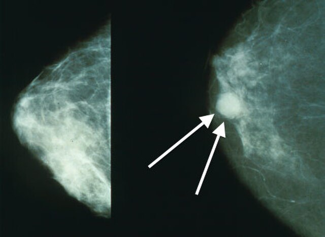
Breast cancer imaging examples are provided in Figures 1 and 2 below.
Figure 18
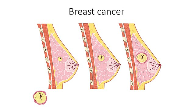
Figure 29
Care Team Timeout Healthcare professionals should be familiar with breast cancer risk factors because early identification and intervention can significantly impact patient outcomes. During wellness or acute visits, PCPs are crucial in assessing a patient’s risk factors, conducting screenings, and guiding patients on preventive measures. They are often a patient’s first point of contact and can initiate referrals to specialists when necessary. Pharmacists can educate patients about risk factors, encourage adherence to screening recommendations, and support the management of medications used for prevention or treatment. Radiologists use mammograms, ultrasounds, and MRIs to detect and evaluate breast abnormalities. Breast care nurses, also called nurse navigators, guide patients through the system and appointments and provide education. |
Clinical Presentation and Diagnosis
Breast cancer can present with a variety of clinical manifestations, depending on the stage and subtype of the disease. Patients are often asymptomatic, and breast cancer is commonly detected through routine mammography screening.2 When symptoms occur, the most common is a painless, palpable lump.2 Less commonly, patients may experience pain, nipple discharge, retraction or dimpling, and skin changes such as edema, redness, or warmth.2 In more advanced stages, breast cancer may present with symptoms related to metastasis, such as bone pain, shortness of breath, jaundice, or neurological symptoms, depending on the site of metastatic spread.2 Approximately 20-30% of women with early-stage breast cancer will eventually develop metastatic breast cancer.2 Accurate and early diagnosis of breast cancer is essential for effective treatment and improved survival outcomes.
The diagnostic approach may involve a combination of a patient’s history, physical examination, imaging, biopsy, and molecular testing.2 Biopsies are vital for confirming a breast cancer diagnosis. Depending on the clinical scenario, it can be performed with fine-needle aspiration, core-needle biopsy, or excisional biopsy.2
Molecular testing of biopsy samples is crucial for determining hormone receptor status, which guides treatment decisions.2 Hormone receptor status includes estrogen receptor and progesterone receptor (ER/PR), as well as Human Epidermal Growth Factor Receptor 2 (HER2) expression. Genetic testing for BRCA mutations and other hereditary cancer syndromes may also be indicated in patients with a strong family history or early-onset breast cancer.2 Diagnostic approaches are summarized in Table 4 below.2
Table 4
Diagnostic Approaches for Breast Cancer Detection2,10
| Diagnostic Tool | Description |
| Mammography |
|
| Ultrasound |
|
| Magnetic Resonance Imaging (MRI) |
|
| Biopsy |
|
| Molecular Testing |
|
In terms of laboratory findings in patients with metastatic breast cancer, tumor markers such as cancer antigen 15-3 (CA 15-3) and carcinoembryonic antigen (CEA) may be elevated.2 Additionally, elevated levels of alkaline phosphatase or abnormal liver function test results can also indicate the presence of metastatic disease.2
Care Team Timeout PCPs are aware of factors that may affect the accuracy of mammography, such as breast density, which can vary with age, menopausal status, and hormone replacement therapy use For women with dense breasts or specific conditions, alternative imaging methods are available, like ultrasonography, MRI, digital mammography, and tomosynthesis Once a diagnosis is confirmed, the PCP collaborates with oncologists, surgeons, and other specialists to develop a comprehensive treatment plan PCPs discuss expectations with patients and approach care plan development in a patient-centric manner Pharmacists can refer patients in need of screening, or when a patient makes the pharmacist aware of alarm symptoms |
Breast Cancer Staging and Prognosis
Breast cancer confined to a localized breast lesion is often referred to as early, primary, localized, or curable.2 When the cancer spreads to local–regional lymph nodes, it is still considered an early stage.2 However, breast cancer cells can spread through lymph channels or the bloodstream to distant sites, leading to advanced or metastatic breast cancer.2 Common sites for distant metastases include lymph nodes beyond the local–regional area, skin, bone, liver, lungs, and brain.2 Symptoms such as bone pain, difficulty breathing, abdominal enlargement, jaundice, and mental status changes may indicate the presence of metastatic breast cancer.2 Some women may present with distant metastases at their initial appointment, often due to a neglected breast mass.2 Additionally, 20% to 30% of patients with early-stage breast cancer will eventually develop metastatic disease.2 The staging of breast cancer is summarized in Table 5.2
Table 5
Breast Cancer Staging2
| TX | The primary tumor cannot be assessed | |
| T0 | No evidence of primary tumor | |
| Tis (DCIS) | Ductal carcinoma in situ | |
| Tis (Paget) | Paget’s disease of the nipple is NOT associated with invasive carcinoma or carcinoma in situ | |
| T1 | Tumor ≤2 cm in greatest dimension | |
| T1mi | <0.1 cm; microinvasion | |
| T1a | >0.1 cm ≤0.5 cm | |
| T1b | >0.5 cm ≤1 cm | |
| T1c | >1 cm ≤2 cm | |
| T2 | Tumor >2 cm ≤5 cm | |
| T3 | Tumor >5 cm | |
| T4 | Tumor of any size with direct extension to the chest wall and/or to the skin | |
| T4a | Extension to the chest wall; invasion or adherence to the pectoralis muscle in the absence of invasion of chest wall structures | |
| T4b | Ulceration and/or ipsilateral macroscopic satellite nodules and/or edema (including peau d’orange) of the skin that does not meet the criteria of inflammatory carcinoma | |
| T4c | Both T4a and T4b present | |
| T4d | Inflammatory carcinoma | |
| Regional Lymph Nodes: Clinical (cN) | ||
| cNX | Regional lymph nodes cannot be assessed | |
| cN0 | No regional lymph node metastases | |
| cN1 | Metastases to movable ipsilateral level 1, II axillary lymph node(s) | |
| cN1mi | Micrometastases (approximately 200 cells, >0.02 cm <0.2 cm) | |
| cN2 | Metastases in ipsilateral level I, II axillary lymph nodes that are clinically fixed or matted; or in ipsilateral internal mammary nodes in the absence of axillary lymph node metastases | |
| cN2a | Metastases in ipsilateral level I, II axillary lymph nodes fixed to one another (matted) or to other structures | |
| cN2b | Metastases only in ipsilateral internal mammary nodes in the absence of axillary lymph node metastases | |
| cN3 | Metastases in ipsilateral infraclavicular (level III axillary) lymph node(s) with or without level I, II axillary lymph node involvement; or in ipsilateral internal mammary lymph node(s) with level I, II axillary lymph node metastases; or metastases in ipsilateral supraclavicular lymph node(s) with or without axillary or internal mammary lymph node involvement | |
| cN3a | Metastases in the ipsilateral infraclavicular lymph node(s) | |
| cN3b | Metastases in ipsilateral internal mammary lymph node(s) and axillary lymph node(s) | |
| cN3c | Metastases in the ipsilateral supraclavicular lymph node(s) | |
| Regional Lymph Nodes: Pathologic (pN) | ||
| pNX | Regional lymph nodes cannot be assessed | |
| pN0 | No regional lymph node metastasis identified or Isolated tumor cells (ITCs) only | |
| pN0(i+) | ITCs only, no larger than 0.02 cm in regional lymph nodes | |
| pN0(mol+) | Positive molecular findings by reverse transcriptase polymerase chain reaction; no ITCs detected | |
| pN1 | Micrometastases; or metastases in 1-3 axillary lymph nodes; and/or clinically negative internal mammary nodes with micrometastases or macrometastases by sentinel lymph node biopsy | |
| pN1mi | Micrometastases, (approximately 200 cells, >0.02 cm <0.2 cm) | |
| pN1a | Metastases in ipsilateral internal mammary sentinel nodes, excluding ITCs | |
| pN1c | pN1a and pN1b combined | |
| pN2 | Metastases in 4-9 axillary lymph nodes; or positive ipsilateral internal mammary lymph nodes by imaging in the absence of axillary lymph node metastases | |
| pN2a | Metastases in 4-9 lymph nodes (at least one deposit >0.2 cm) | |
| pN2b | Metastases in clinically detected internal mammary lymph nodes with or without microscopic confirmation; with pathologically negative axillary nodes | |
| pN3 | Metastases ≥10 or more axillary lymph nodes; or in infraclavicular (level III axillary) lymph nodes; or positive ipsilateral internal mammary lymph nodes by imaging in the presence ≥1 positive level I, II axillary lymph nodes; or in >3 axillary lymph nodes and micrometastases or macrometastases by sentinel lymph node biopsy in clinically negative ipsilateral internal mammary lymph nodes; or in ipsilateral mammary lymph nodes | |
| pN3a | Metastases in ≥10 axillary lymph nodes (at least one tumor deposit >0.2 cm) or metastases to infraclavicular (level III axillary lymph) nodes | |
| pN3b | pN1a or pN2a in the presence of cN2b (positive internal mammary nodes by imaging) or pN2a in presence of pN1b | |
| pN3c | Metastases in the ipsilateral supraclavicular lymph nodes | |
| Distant Metastasis (M) | ||
| M0 | No clinical or radiographic evidence of distant metastases | |
| cM0(i+) | No clinical or radiographic evidence of distant metastases in the presence of tumor cells or deposits <0.02 cm detected microscopically or by molecular techniques in circulating blood, bone marrow, or other nonregional nodal tissue in a patient without symptoms or signs of metastases | |
| cM1 | Distant metastases detected by clinical and radiographic means | |
| pM1 | Any histologically proven metastases in distant organs; or if in nonregional nodes, metastases >0.02 cm | |
How breast cancer progresses is incredibly patient-specific. Some patients may experience a rapidly advancing and aggressive form of the disease, while others have a slower course.2 In determining strategic, personalized treatment plans for patients, clinicians need to be able to predict prognoses accurately.2 Prognostic factors are characteristics or measurements that become available at diagnosis or during surgery and are associated with outcomes like recurrence or survival rates independent of any additional systemic therapy.2 Predictive factors, on the other hand, help determine the likelihood of a patient’s response to specific treatments.2 These factors encompass a range of categories, including patient characteristics such as age, cancer-specific features like tumor size and histologic type, biomarkers such as hormone-receptor status, and genetic variables.2
Prognostic and predictive factors for breast cancer outcomes include both patient and cancer characteristics as well as biomarkers.2 Younger age (under 35) and African American race are associated with a worse prognosis. Key cancer characteristics influencing prognosis are tumor size, lymph node involvement, or lymphovascular invasion, nuclear grade (with grade 3 being the most aggressive), and histologic subtype, particularly invasive ductal carcinoma.2 Biomarkers also play a significant role, with estrogen and progesterone receptor positivity (ER/PR) being favorable, while HER2 positivity is generally associated with a poorer prognosis.2
Treatment Modalities
The treatment of breast cancer is multidisciplinary and may involve surgery, radiation therapy, systemic therapy, and supportive care. The treatment approach depends on the disease stage and patient factors, including age, comorbidities, and preferences. The goal of treatment varies by stage: in early-stage breast cancer, the primary objective is curative, while in advanced metastatic breast cancer, the focus may shift toward palliation and quality of life. Early-stage or curative breast cancer often involves the use of various treatment strategies, including surgery, radiation, and systemic medication therapies.2
The management of metastatic breast cancer promotes disease regression and optimizes a patient’s quality of life.2 The approach is personalized based on a tumor’s characteristics, prior treatments, and the patient's overall health and preferences.2
Care Team Timeout Pathologists examine biopsied tissue samples and cells to determine the type and characteristics of the cancer. Once a diagnosis is made, PCPs coordinate care, manage comorbid conditions, and address the overall health of the patient Monitoring treatment side effects, managing chronic conditions, and ensuring patients adhere to prescribed therapies contribute to outcomes. Ongoing review of the patient's comorbidities, current medications, and treatment history enables Identification of potential drug interactions and the need for dose adjustments Assessing a patient's readiness to follow the treatment plan and manage any side effects or complications from prior therapies is important throughout the patient’s journey through their diagnosis and treatment. |
Surgery
The management of early invasive breast cancer treatment may include breast-conserving surgery (BCS) followed by radiotherapy (RT) and mastectomy with or without reconstruction.1 BCS involves removing part of the breast with cancer and surrounding margins, while mastectomy involves the removal of the entire breast.11 BCS combined with RT offers similar local recurrence rates (LRR) and survival outcomes compared to mastectomy, particularly for estrogen receptor (ER) positive and HER2-positive breast cancers.1 It is often preferred due to better cosmetic and patient satisfaction outcomes.1 BCS may not be appropriate for patients with extensive micro-calcifications or those unable to achieve negative margins.1 Neoadjuvant chemotherapy (NAC) may be used to shrink the tumor and facilitate BCS for patients with large tumors.1 The management of axillary lymph nodes varies based on the initial status and the use of neoadjuvant therapy, with sentinel lymph node (SLN) biopsy often sufficient for clinically node-negative patients.1 Surgery is also crucial in managing local-regional recurrences, with mastectomy or wide resection being standard, depending on previous treatments.1
Chemotherapy
The use of chemotherapy in breast cancer includes neoadjuvant (NAC), adjuvant (AC), and salvage therapies, which may significantly reduce the risk of recurrence.1 Neoadjuvant therapy is given before surgery to shrink the tumor’s size and increase the effectiveness of the surgery.1 AC is given after surgery to kill the remaining cancer cells and prevent the recurrence of the cancer.1 Salvage therapy is used once other treatments have failed.1 NAC is particularly beneficial for downstaging tumors, making them operable, or facilitating breast conservation surgery.1 It is recommended for large tumors, multiple lymph node involvement, and aggressive subtypes like triple-negative and HER2-positive cancers.1 NAC allows for the assessment of treatment response and guides subsequent therapies.1
In the treatment of early-stage cancer, chemotherapy regimens can be anthracycline-based or non-anthracycline-based.1 The anthracycline-based regimen typically involves doxorubicin and cyclophosphamide administered every 2-3 weeks for four cycles, followed by a taxane, such as weekly paclitaxel for 12 weeks or docetaxel every three weeks for four cycles.1 The non-anthracycline option includes docetaxel and cyclophosphamide every three weeks for four cycles.1
Adjuvant chemotherapy is typically administered within three months post-surgery, with dose-dense regimens (every 14 days) showing better outcomes than traditional schedules.1 Emerging evidence supports specific regimens, such as adding platinum for triple-negative breast cancer or using capecitabine maintenance therapy for improved survival.1
Radiotherapy
Radiotherapy (RT) remains a cornerstone of treatment after breast-conserving surgery (BCS), particularly with whole breast irradiation (WBI) or hypofractionated WBI for early-stage breast cancer.1 For selected low-risk patients, partial breast irradiation (PBI) or accelerated PBI may be viable alternatives, offering similar outcomes with reduced toxicity.1 Post-mastectomy RT (PMRT) is standard for patients with four or more positive axillary lymph nodes, although its benefit in those with one to three nodes is still under study.1 Regional nodal irradiation, including the chest wall and supraclavicular areas, improves disease-free survival, especially involving internal mammary nodes (IMNs).1 However, the latter remains controversial due to potential increased toxicity.1
Endocrine Therapy
Endocrine therapy is a standard adjuvant treatment for hormone receptor-positive (HR+) breast cancer, and the duration of endocrine therapy is typically 5 to 10 years.1 In premenopausal women, tamoxifen 20 mg per day is commonly used, with extended treatment of up to 10 years showing further reductions in recurrence and mortality.1 For high-risk patients, combining ovarian suppression with an aromatase inhibitor (AI) and tamoxifen improves outcomes, though with higher toxicity.1 In postmenopausal women, AIs are often preferred due to better efficacy, particularly in high-risk cases, though they are associated with higher bone-related side effects.1 The duration and combination of endocrine therapies may be guided by multigene assays, particularly in endocrine-resistant cases, where CDK inhibitors may be added for better outcomes.1
Targeted Therapy
Targeted therapy, particularly for HER2-positive breast cancer, has revolutionized treatment, with trastuzumab (an anti-HER2 drug) being the cornerstone.1 Combining trastuzumab with chemotherapy significantly reduces recurrences and improves survival, making it a standard in neoadjuvant and adjuvant settings.1 Additional anti-HER2 agents like pertuzumab and lapatinib have shown benefits in increasing pathological complete response (pCR) rates and improving outcomes in HER2-positive cancers.1 Early use of anti-HER2 therapies such as docetaxel with trastuzumab and pertuzumab is recommended for metastatic disease, with newer agents like trastuzumab emtansine (TDM-1) offering benefits in previously treated patients.1
Immunotherapy
Breast cancer's ability to evade immune responses is partly due to inhibitory interactions between T-cell receptors (such as PD-1, TIM-3, LAG3, TIGIT, and CTLA4) and their ligands on cancer cells, leading to T-cell exhaustion and reduced therapy responsiveness.1 Single-agent immune checkpoint inhibitors (ICIs) have shown limited effectiveness in advanced breast cancer, particularly in metastatic triple-negative breast cancer (TNBC), with modest response rates observed in trials like KEYNOTE-012, KEYNOTE-086, and JAVELIN.1 However, combining ICIs with chemotherapy has yielded better outcomes, as seen in trials like IMpassion130 and KEYNOTE-522, leading to FDA approval of atezolizumab and pembrolizumab for first-line treatment in PD-L1-positive metastatic TNBC.1 In early-stage breast cancer, the addition of ICIs to neoadjuvant chemotherapy has shown promise, especially in TNBC, though further research is needed to confirm long-term efficacy.1
Gene Therapy
Gene therapy is an emerging approach to breast cancer treatment that involves delivering genetic material to target cells to modify gene expression.1 Early trials have demonstrated safety and targeted gene expression in breast cancer, particularly in HER2-positive cases.1 Strategies include gene editing, microRNA therapy, and DNA/RNA vaccination.1 Although still in the experimental phase, gene therapy holds promise for the future, with ongoing trials exploring its potential in treating breast cancer
Care Team Timeout Subsequent to discharge from the surgeon, radiation oncologist and/or plastic surgeon, social workers are included in patient care, and PCPs monitor for signs of recurrence, manage any long-term side effects of treatment, and coordinate care with other specialists, as needed. They also play a crucial role in providing psychosocial support and addressing the emotional and psychological needs of breast cancer patients, which is an essential aspect of holistic care. Communication and collaboration remain key to complex patient care. Pharmacists utilize their expertise in drug interactions and adverse effects management to optimize patient outcomes following the initiation of therapies They work closely with the care team to recommend therapeutic changes as needed and monitor a patient’s adherence to therapy Pharmacists in community settings can provide cancer education, leading to better patient attitudes toward the disease, improved adherence to treatment, and more effective pain management |
Care Team Barriers
Care team barriers to interprofessional collaborative practice include communication gaps, time constraints, keeping up with rapidly evolving treatments, limited patient adherence to treatment plans, navigating complex insurance and financial issues, and managing side effects and complications. These barriers, challenges, and solutions are described in Table 6 below.
Table 6
Care Team Barriers
| Barrier | Challenges | Solutions |
| Communication Gaps | Fragmented care due to poor interdisciplinary communication | - Regular interdisciplinary meetings or case conferences - Integrated electronic health records (EHR) systems for seamless information sharing |
| Time Constraints | Limited time for thorough patient evaluation and management | - Delegate non-clinical tasks to support staff - Use automated systems for routine processes - Prioritize care activities based on patient acuity |
| Keeping Up with Rapidly Evolving Treatments | Difficulty staying current with new drugs and therapies | - Access to continuing education (online courses, webinars, conferences) - Decision support tools within EHRs for evidence-based recommendations |
| Limited Patient Adherence to Treatment | Non-adherence due to side effects, financial constraints, or lack of understanding | - Enhance patient education on adherence and side effect management - Identify and address barriers to adherence (e.g., financial assistance) |
| Navigating Complex Insurance and Financial Issues | Complications from insurance limitations and high treatment costs | - Collaborate with financial counselors and patient navigators - Advocate for clear communication with insurance companies |
| Managing Side Effects and Complications | Challenges in managing side effects like cardiotoxicity, neuropathy, and bone marrow suppression | - Establish early identification and management protocols - Use a multidisciplinary approach involving specialists as needed |
Patient Case
A 58-year-old woman presents to the clinic after noticing a firm, painless lump in her left breast.
What patient history questions should be asked?
The patient’s history reveals menarche at age 11, first childbirth at age 34, and her mother was diagnosed with breast cancer in her early 50s. She is postmenopausal, with a BMI of 31 kg/m2, and she drinks alcohol several times per week. Her last screening mammogram was three years ago.
On exam, a 2.5 cm irregular mass is palpated in the upper outer quadrant of the left breast with a slightly enlarged ipsilateral axillary lymph node. Diagnostic mammography and targeted ultrasound are ordered, followed by core needle biopsy. The biopsy reveals estrogen receptor-positive invasive ductal carcinoma that is also progesterone receptor positive and HER2 negative.
What is the next step for this patient?
The care team understands the importance of patient support and compassionate, personalized care. The patient is first provided with support tools, including 24/7 helplines, peer mentorship, and resource navigation within the Susan G. Komen organization and the American Cancer Society. The patient is referred to the breast surgery team, medical oncology, radiation oncology, pathology, and pharmacy for coordinated treatment planning. The interdisciplinary team discusses surgery, adjuvant endocrine treatment, and surveillance while addressing symptom management, adherence, and psychosocial support.
Guidelines and Resources
| American Cancer Society (ACS) |
| National Comprehensive Cancer Network (NCCN) Guidelines for Patients |
| American Society of Clinical Oncology (ASCO) |
| European Society for Medical Oncology (ESMO) |
| St. Gallen International Expert Consensus |
| United States Preventive Services Task Force (USPSTF) |
| American College of Radiology (ACR) Appropriateness Criteria |
| Society of Surgical Oncology (SSO) |
| San Antonio Breast Cancer Symposium (SABCS) Consensus |
| American College of Surgeons (ACS) Commission on Cancer (CoC) |
Summary
Breast cancer management requires a coordinated multidisciplinary approach to achieve the best patient outcomes. Physicians and pharmacists, along with other healthcare professionals, must work together to provide comprehensive care that addresses the complex needs of breast cancer patients. Through continuing education and collaboration, healthcare professionals can help advance breast cancer care and improve patient survival and quality of life. As treatment strategies continue to evolve, the roles of pharmacists and primary care physicians in the management of breast cancer will remain critical in ensuring that patients receive the best possible care throughout their journey.
References
Wang J, Wu SG. Breast Cancer: An Overview of Current Therapeutic Strategies, Challenge, and Perspectives. Breast Cancer (Dove Med Press). 2023;15:721-730. Published 2023 Oct 20. doi:10.2147/BCTT.S432526.
Boster B, Patel NK, Kaushik J. Breast Cancer. In: DiPiro JT, Yee GC, Haines ST, Nolin TD, Ellingrod VL, Posey L. eds. DiPiro’s Pharmacotherapy: A Pathophysiologic Approach, 12th Edition. McGraw Hill; 2023.
Testa U, Castelli G, Pelosi E. Breast Cancer: A Molecularly Heterogenous Disease Needing Subtype-Specific Treatments. Med Sci (Basel). 2020;8(1):18. Published 2020 Mar 23. doi:10.3390/medsci8010018
Łukasiewicz S, Czeczelewski M, Forma A, Baj J, Sitarz R, Stanisławek A. Breast Cancer-Epidemiology, Risk Factors, Classification, Prognostic Markers, and Current Treatment Strategies-An Updated Review. Cancers (Basel). 2021;13(17):4287. Published 2021 Aug 25. doi:10.3390/cancers13174287
National Cancer Institute. Breast Cancer Risk Assessment Tool: Online Calculator (The Gail Model). National Cancer Institute. Undated. https://bcrisktool.cancer.gov/. Accessed October 14, 2024.
American Cancer Society. Recommendations for the Early Detection of Breast Cancer. ACS. December 19, 2023. https://www.cancer.org/cancer/types/breast-cancer/screening-tests-and-early-detection/american-cancer-society-recommendations-for-the-early-detection-of-breast-cancer.html#:~:text=Women%20between%2040%20and%2044,choose%20to%20continue%20yearly%20mammograms. Accessed October 14, 2024.
US Preventive Services Task Force. Breast Cancer: Screening. USPSTF. April 30, 2024. https://www.uspreventiveservicestaskforce.org/uspstf/recommendation/breast-cancer-screening#:~:text=What%20is%20the%20Task%20Force,of%20dying%20from%20this%20disease. Accessed October 14, 2024.
Wikicommons. Breast Cancer. Wikimedia.org. https://commons.wikimedia.org/wiki/File:Breast_cancer_--_Smart-Servier.jpg. Accessed October 14, 2024.
Wikicommons. Mammo Breast Cancer. https://commons.wikimedia.org/wiki/File:Mammo_breast_cancer_wArrows.jpg. Accessed October 14, 2024.
Basurto-Hurtado JA, Cruz-Albarran IA, Toledano-Ayala M, Ibarra-Manzano MA, Morales-Hernandez LA, Perez-Ramirez CA. Diagnostic Strategies for Breast Cancer Detection: From Image Generation to Classification Strategies Using Artificial Intelligence Algorithms. Cancers (Basel). 2022;14(14):3442. Published 2022 Jul 15. doi:10.3390/cancers14143442
ACS. Masectomy. December 20, 2023. https://www.cancer.org/cancer/types/breast-cancer/treatment/surgery-for-breast-cancer/mastectomy.html. Accessed October 14, 2024.
ASHP-HOPA Guidelines on the Roles and Responsibilities of the Pharmacy Technician in Ambulatory Oncology Pharmacy. 2018. https://www.ashp.org/-/media/assets/policy-guidelines/docs/guidelines/pharmacy-technician-role-responsibilities-ambulatory-oncology-pharmacy.pdf. Accessed October 14, 2024.
DISCLAIMER
The information provided in this course is general in nature, and it is designed solely to provide participants with continuing education credit(s). This course and materials are not meant to substitute for the independent, professional judgment of any participant regarding that participant’s professional practice, including but not limited to patient assessment, diagnosis, treatment, and/or health management. Medical and pharmacy practices, rules, and laws vary from state to state, and this course does not cover the laws of each state; therefore, participants must consult the laws of their state as they relate to their professional practice.
Healthcare professionals must consult their employer, healthcare facility, hospital, or other organization for guidelines, protocols, and procedures to follow. The information provided in this course does not replace those guidelines, protocols, and procedures, but is for academic purposes only, and this course’s limited purpose is for the completion of continuing education credits.
Participants are advised and acknowledge that information related to medications, their administration, dosing, contraindications, adverse reactions, interactions, warnings, precautions, or accepted uses is constantly changing. Any person taking this course understands that such a person must make an independent review of medication information before any patient assessment, diagnosis, treatment and/or health management. Any discussion of off-label use of any medication, device, or procedure is informational only, and such uses are not endorsed hereby.
Nothing contained in this course represents the opinions, views, judgments, or conclusions of RxCe.com LLC. RxCe.com LLC is not liable or responsible to any person for any inaccuracy, error, or omission with respect to this course or course material.
© RxCe.com LLC 2026: All rights reserved. No reproduction of all or part of any content herein is allowed without the prior, written permission of RxCe.com LLC.
RxCe.com
© RxCe.com LLC 2025: All rights reserved.